Efficient gene delivery is essential for advancing cell and gene therapies. Polyethylenimine (PEI) transfection has become a widely adopted method due to its high DNA delivery efficiency, cost-effectiveness, and compatibility with multiple cell lines. Unlike lipid-based reagents, PEI offers scalability from early-stage research to large-scale commercial manufacturing, making it a practical choice for both discovery and production workflows.
Achieving consistent transfection efficiency requires optimization of multiple interdependent variables: DNA:PEI ratio, cell density, media composition, complexation conditions, and timing. Small deviations in any of these parameters can significantly affect titers or cause cytotoxicity. This guide provides a structured approach to PEI protocol optimization, with specific recommendations for maximizing efficiency while minimizing toxicity.
Understanding PEI Transfection
PEI-mediated transfection relies on electrostatic interactions between the positively charged polymer and the negatively charged DNA backbone. As a polycation, PEI condenses DNA into compact polyplexes that protect against nuclease degradation and enable cellular uptake via endocytosis. Once internalized, the protonatable amines on PEI facilitate endosomal escape through the "proton sponge" effect, releasing DNA into the cytoplasm for nuclear entry and expression.
PEI architecture and molecular weight
PEI is available in linear and branched forms, each with distinct performance characteristics. Linear PEI forms more uniform complexes and generally exhibits lower cytotoxicity than branched PEI at equivalent molecular weights. Among linear variants, 25 kDa PEI has emerged as the industry standard as it balances high transfection efficiency with acceptable cytotoxicity [1].
Lower-molecular-weight PEI (e.g., 10 kDa) forms less stable polyplexes and thus have lower transfection efficiency. Higher-molecular-weight or branched PEI increases complex stability but often at the cost of elevated cytotoxicity and greater batch-to-batch variability [2, 3]. For most applications, 25 kDa linear PEI offers the best compromise between performance and cell health.
Key variables affecting transfection efficiency
Multiple interdependent factors determine whether a transfection succeeds or fails:
-
Cell type and state - Different lines have distinct uptake capacities and sensitivities to PEI. Passage number, growth phase, confluency (adherent) or density (suspension), and viability all affect performance.
-
DNA quality and purity - Endotoxin contamination, residual protein, and degraded DNA reduce efficiency and increase toxicity.
-
N/P ratio - The balance of protonatable amines in PEI to phosphate groups in DNA sets the net charge of the complex, which governs aggregation, uptake, and endosomal escape.
-
DNA concentration -Total DNA per cell must be optimized independently of the N/P ratio.
-
PEI quality and age - Degraded or improperly stored PEI loses charge density, affecting DNA condensation and ultimately transfection efficiency.

Dos and Don'ts for High-Efficiency PEI Transfection
High-efficiency transfection requires disciplined protocol execution. The recommendations below address the most common sources of variability and provide a framework for systematic optimization.
DO: Optimize DNA:PEI Ratio
The N/P ratio is the single most critical parameter in PEI transfection. It defines the net charge of the polyplex, which controls aggregation, cellular uptake, endosomal escape, and cytotoxicity. Every cell line has a distinct optimal N/P range, typically between 5 and 20 for 25 kDa linear PEI. Optimization requires systematic titration.
Initial DNA amount. Begin with 0.5 to 3 µg of plasmid DNA per 1 × 10^6 cells. This range works for most mammalian cell types, but the optimal amount depends on factors such as plasmid size, promoter strength, and the number of plasmids that are co-transfected.
Run a dose-response matrix. Test a grid of DNA:PEI ratios (or N/P ratios). Measure both expression level and cell viability at 24, 48, and 72 hours post-transfection. The goal is to identify the minimal DNA and PEI input that delivers maximal expression without compromising viability.
Include essential controls:
-
Positive control - A validated expression plasmid (e.g., CMV-GFP) confirms that the transfection reagent and cells are performing as expected.
-
Negative controls - PEI-only and DNA-only controls establish baseline fluorescence or luminescence.
Use reporters during optimization. GFP provides a rapid percentage-positive readout via flow cytometry or microscopy. Luciferase offers high dynamic range and quantitative output suitable for plate-reader assays. Once optimal conditions are established with a reporter, validate them with your production construct before scaling.
Account for plasmid-specific variables. Large plasmids (>10 kb), weak promoters, or co-transfection of multiple plasmids may require higher DNA input or adjusted N/P ratios. Re-optimize when switching constructs or introducing new plasmids to the workflow.
For AAV and lentiviral vector production, complete N/P optimization with a reporter system first, then re-verify conditions under production settings before committing to scale-up.
DO: Use High-Quality, Endotoxin-Free DNA
DNA purity directly impacts transfection efficiency and cell viability. Endotoxin contamination activates innate immune responses, reduces transgene expression, and increases cytotoxicity. Even low levels of endotoxin (>1 EU/µg DNA) can degrade performance in sensitive cell lines.
Quality metrics to monitor:
-
A260/280 ratio - Target 1.8 to 2.0 for pure DNA. Ratios below 1.8 indicate protein contamination; ratios above 2.0 suggest RNA contamination.
-
Endotoxin level - Measure by LAL assay. For research applications, <10 EU/µg is acceptable; for vector production or sensitive cell lines, aim for <1 EU/µg.
-
Visual inspection - High-quality DNA should appear clear in solution. Cloudiness or stringiness indicates degradation or impurities.
Use endotoxin-free plasmid preparation kits or commercial-grade plasmids for critical applications. If endotoxin levels are elevated, endotoxin removal kits can be used to reduce contamination. Always verify endotoxin content after purification.
DO: Maintain Healthy Cells
Cell health determines transfection success. Stressed, over-passaged, or contaminated cells exhibit reduced uptake, lower expression, and higher susceptibility to PEI-mediated toxicity.
Passage number - Genetic drift accumulates with extended passaging, reducing transfection efficiency and altering phenotype. Use cells at passage 5-20, or within 4 to 5 passages post-thaw from a qualified cell bank.
Growth phase - Transfect cells during active growth (log phase). Cells in lag phase or stationary phase have reduced metabolic activity and lower DNA uptake.
Confluency and density - For adherent cells, seed to reach 85 to 90% confluency on the day of transfection. Over-confluent cultures undergo contact inhibition and resist transfection; sparse cultures have reduced viability. For suspension cells, maintain a density of approximately 2 × 10^6 cells/mL at the time of complex addition.
Viability - Ensure ≥95% viability before transfection. Compromised cultures amplify toxicity and produce inconsistent results. Assess viability using a hemocytometer, an automated cell counting machine, such as a Vi-Cell® Blue, or flow cytometry.
Post-transfection monitoring - Track viability at 24, 48, and 72 hours post-transfection using an automated cell counter or a cell viability assay. A drop in viability below 80% generally indicates excessive cytotoxicity, which requires further assay optimization..
DO: Store PEI Solutions Correctly
PEI stability degrades over time, particularly when subjected to repeated freeze-thaw cycles. Degraded PEI loses charge density, forms inconsistent complexes, and produces variable transfection efficiency.
Storage recommendations:
-
For powders, store at room temperature in a tightly sealed container. For solutions and stock solutions prepared from powders, prepare working aliquots immediately upon receipt to avoid contamination.
-
Store aliquots at the temperature recommended by the manufacturer (typically 4℃ for working stocks for PEI hydrochloride).
-
Date each aliquot and discard after the recommended shelf life.
-
Verify PEI performance periodically with a standard transfection control.
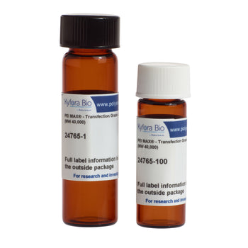
PEI MAX® powder from Kyfora Bio is stable at room temperature until expiration. PEI MAX® stock solution is stable for 6 to 12 months when stored at 4℃. Transporter 5® transfection reagent, crafted from the PEI MAX® powder at a 1mg/mL concentration, is stable at 4℃ until expiration. Due to the long storage capacity of our reagents at 4℃, the need to freeze aliquots to extend lifetime is eliminated.
DO: Optimize DNA-PEI Complexation Time
Complex formation requires time for electrostatic interactions to stabilize. Incubation periods that are too short yield incomplete complexes with poor uptake; incubation periods that are too long promote aggregation and increase cytotoxicity.
Standard complexation time: 10 to 30 minutes at room temperature is appropriate for most applications. The optimal time depends on factors including plasmid size, DNA concentration, PEI molecular weight, and buffer composition.
Signs of suboptimal timing:
-
Too short - Low or variable expression, visible incomplete mixing
-
Too long - Large aggregates visible by microscopy, reduced viability, lower expression
Once optimized, hold complexation time constant across all experiments to reduce variability.
DO: Optimize Transfection Timing and Harvest Window
Expression kinetics vary by construct, promoter, cell line, and transfection efficiency. Identifying the optimal harvest window requires empirical testing.
Timing of complex addition: For adherent cells, add complexes 18 to 24 hours after seeding, aiming for a confluency of 85 to 90%. For suspension cells, add complexes when cultures are in mid-log phase at approximately 2 × 10^6 cells/mL with ≥95% viability.
Harvest window determination: Peak transgene expression typically occurs between 48 and 96 hours post-transfection, but varies depending on a variety of factors. Run a time-course experiment measuring expression at 24, 48, 72, and 96 hours.
Signs of suboptimal harvest timing:
-
Too early - Low yield, insufficient accumulation
-
Too late - Protein degradation, reduced viability, cell detachment (adherent), vector loss due to cell lysis
Once the optimal window is identified, lock the harvest time for all production runs. Monitor cell viability alongside expression to ensure the culture remains healthy throughout the production window.
DON'T: Allow Cells to Become Overconfluent
Confluency and cell density are critical determinants of transfection success. Both over- and under-confluent cultures perform poorly.
Adherent cells - Overconfluence triggers contact inhibition, reducing metabolic activity and DNA uptake. Cultures below 70% confluency exhibit lower viability and reduced transfection efficiency. Seed cells 18 to 24 hours before transfection to reach 85 to 90% confluency on the day of complex addition. Some robust lines tolerate up to 95% confluency, but this should be verified empirically.
Suspension cells - Maintain mid-log phase density at the time of transfection. Target approximately 2 × 10^6 cells/mL with ≥95% viability. Overgrown cultures (>3 × 10^6 cells/mL) deplete nutrients, accumulate toxic metabolites, and exhibit reduced transfection efficiency. Very low densities (<5 × 10^5 cells/mL) may not support efficient complex uptake or sustained expression.
DON'T: Use Compromised Cells
Cell contamination with mycoplasma or bacteria can be hard to diagnose as a cause of failure in transfection assays. Infected cells exhibit altered metabolism, disrupted signal transduction, and reduced DNA uptake. Even low-level contamination degrades reproducibility and makes optimization impossible.
Impact of bacteria and mycoplasma contamination
-
Reduced transfection efficiency (often by 50% or more)
-
Increased cytotoxicity
-
Altered gene expression profiles
-
Inconsistent results across experiments
Prevention and detection: Test all cell lines quarterly using PCR-based mycoplasma detection kits. Maintain aseptic technique, avoid sharing media or reagents between cultures, and quarantine new cell lines until testing confirms they are bacteria and mycoplasma-free. Use antibiotics and antimycotics for culturing cells, but not when seeding for transfection. Discard contaminated cultures immediately—decontamination is unreliable and risks cross-contamination.
DON'T: Use Serum, Antibiotics, or Antimycotics During Complex Formation
Serum proteins can interfere with PEI-DNA complex formation and stability. Antibiotics and antimycotics can be taken up by cells during endocytosis of the PEI-DNA complex, causing toxicity. In both instances, the end result is a decrease in transfection efficiency. Complexation buffer requirements:
-
Serum-free - Serum proteins coat complexes and block cellular uptake
-
Antibiotic-free, Antimycotic-free - Antibiotics and antimycotics can enter the cells during endocytosis of the PEI/DNA complex
-
Avoid phosphate buffers - Phosphate competes with DNA for PEI binding
Form complexes in buffers compatible with transfection. After complex addition, maintain serum-free, antibiotic-free conditions for 4 to 6 hours to allow efficient uptake. Return to complete medium if the cell line requires serum for long-term viability.
Commonly Used Transfection Complex Formation Media
|
Medium |
Description |
|
Opti-MEM™ |
A reduced-serum medium designed for the growth of a wide range of mammalian cells, often used for transfection due to its low serum content and compatibility with various transfection reagents. |
|
OptiPro™ |
A serum-free, protein-free medium specifically formulated for the growth of a wide variety of cell types, making it suitable for transfection where serum interference is a concern. |
|
DMEM (without supplements) |
Dulbecco's Modified Eagle Medium, a basal medium often used for complex formation when serum and other supplements are omitted to avoid interference with transfection. |
|
RPMI-1640 (without supplements) |
A basal medium originally developed for suspension cultures of human leukemic cells, widely used for transfection of lymphocytes, hybridomas, and other mammalian cell types when serum and supplements are omitted to prevent interference with transfection reagent complexes. |
|
DMEM/F-12 (without supplements) |
A 1:1 mixture of Dulbecco's Modified Eagle Medium and Ham's F-12 Nutrient Mixture that combines the benefits of both formulations, commonly used for transfection of a broad range of adherent cell types when prepared without serum or supplements to ensure efficient complex formation. |
|
Culture media without supplements |
Any basal cell culture medium used without the addition of serum, antibiotics, or other supplements to ensure optimal complex formation and reduce toxicity. |
DON'T: Neglect Post-Transfection Care
Residual free PEI and unbound complexes remaining in the culture medium can increase cytotoxicity, particularly in sensitive cell lines. Media replacement 4 to 6 hours post-transfection removes excess PEI and supports cell recovery.
When to replace media:
-
For sensitive cell lines or high N/P ratios (>15), replace media 4 to 6 hours post-transfection
-
For robust cell lines at moderate N/P ratios (5 to 10), media replacement may be optional
-
Test both conditions empirically and compare viability and expression at 48 hours
Simplified workflows: Kyfora Bio's PEI formulations are optimized to minimize cytotoxicity, often eliminating the need for media replacement and simplifying production workflows.
Troubleshooting
Low transfection efficiency/low or no titers
Possible causes: Poor DNA quality, suboptimal N/P ratio, incorrect complexation conditions, low cell viability, inactive promoter
Solutions:
-
Verify DNA quality: A260/280 ratio of 1.8 to 2.0, endotoxin <1 EU/µg, no visible degradation
-
Confirm plasmid sequence and promoter activity in the target cell line
-
Optimize PEI:DNA ratio using a dose-response curve.
-
Optimize DNA amount per cell using a dose response curve.
-
Check cell passage number, viability (≥95%), and confluence/density.
High cytotoxicity
Possible causes: Excessive PEI, high N/P ratio compromised cell health, contamination
Solutions:
-
Optimize PEI:DNA ratio using a dose-response curve.
-
Increase N/P ratio in increments of 2 to 5 (e.g., test N/P 5, 10, 15, 20)
-
Increase DNA amount per cell using a dose response curve. (test 1, 2, 3 µg per 10^6 cells)
-
Verify cells are mycoplasma-free and healthy
-
Verify cells are in log phase with ≥95% viability at transfection
-
For adherent cells, confirm 85 to 90% confluency; for suspension, 2 × 10^6 cells/mL
-
Test DNA for endotoxin; use endotoxin removal kit if needed
-
Verify PEI storage conditions and age
Variable results between experiments
Possible causes: Inconsistent cell state, reagent degradation, uncontrolled timing, poorly documented protocol
Solutions:
-
Standardize passage number, seeding density, and time from seeding to transfection
-
Prepare fresh PEI and DNA stocks; aliquot to prevent contamination. Standardize complexation time (e.g., always 15 minutes at room temperature)
-
Record all experimental parameters in a protocol checklist
-
Use the same lot of cells, media, and reagents across optimization experiments
Conclusion
Consistent and efficient gene delivery depends on careful optimization of each step in the PEI transfection protocol. Key factors—the DNA:PEI ratio, DNA purity, cell health, complexation chemistry, and timing—directly influence transfection efficiency and reproducibility.
A systematic optimization approach, starting with N/P ratio determination and followed by refinement of cell state, media conditions, and timing parameters, establishes a robust protocol that scales from discovery to manufacturing. When these variables are controlled, PEI transfection delivers reliable performance for gene editing workflows, protein production, and vector manufacturing.
How Kyfora Bio Supports Transfection Optimization
Kyfora Bio, backed by Polysciences' 60+ years of polymer chemistry expertise, offers high-performance PEI transfection reagents designed for the production of viral vectors (AAV and LV), monoclonal antibodies, and recombinant proteins. Our RUO reagents, PEI MAX® and Transporter 5® deliver high transfection efficiency across diverse cell types, reduced cytotoxicity, and cost efficiency compared to competitor reagents. Our GMP grade transfection reagent, MAXgene®, combines the proven performance of our research-grade products with the scalability, validation, and regulatory compliance needed for a smooth transition from early-stage discovery through clinical trials and commercialization.
Our technical team of highly skilled scientists provides protocol optimization support, troubleshooting guidance, and complete documentation packages for regulatory filings.
Request samples or technical consultation. Contact our team to discuss your transfection challenges or request PEI MAX® samples for your workflow.
References:
Boussif O, Lezoualc’h F, Zanta MA, et al. A versatile vector for gene and oligonucleotide transfer into cells in culture and in vivo: polyethylenimine. Proc Natl Acad Sci U S A. 1995;92(16):7297–7301.
Kafil V, Omidi Y. Cytotoxic impacts of linear and branched polyethylenimine nanostructures in A431 cells. Bioimpacts. 2011;1(1):23–30.
Kunath K, Von Harpe A, Fischer D, et al. Low-molecular-weight polyethylenimine as a non-viral vector for DNA delivery: comparison of physicochemical properties, transfection efficiency and in vivo distribution with high-molecular-weight polyethylenimine. J Control Release. 2003;89(1):113–125.